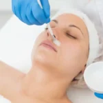
The eyes are often one of the first areas of the face to show signs of aging, due to the delicate skin in this region. Loss of elasticity in the eyelid may result in what is commonly referred to as “hooding,” where excess, loose upper eyelid skin droops outwards and down onto the eyelashes. Age-related changes can also cause the orbital fat, especially the medial fat pad of the upper eyelid, to prolapse through the thin attenuated orbital septum, creating an unsightly bulge. Overall, this can make a person look tired and aged, and is a cause for concern in many patients.Apart from the aging process, eyelid hooding can also occur if the eyelid tissue periodically swells (medically known as blepharochalasis), or with other medical conditions, such as Ehlers-Danlos syndrome, thyroid eye disease, amyloidosis, eye trauma, hereditary angioneurotic edema, elastolysis, renal failure, and xanthelasma.
Symptoms of dermatochalasis
Medical vs cosmetic
Patients may request to undergo surgery to correct upper eyelid dermatochalasis for a myriad of reasons. Chiefly, the goal is to correct the aged appearance that this condition can give. Dermatochalasis can also create a feeling of heaviness in the eyelids and can affect the visual field if the condition is severe, particularly with lateral hooding.
Eyelid hooding may perturb the normal upper eyelid pretarsal show, which can affect eye makeup application. In some patients, mascara applied on the upper lid lashes may run due to contact with the drooping skin. Watery eyes can also result from irritation caused by the shedding of dead skin cells and debris into the eyes. Lateral hooding may cause tearing at the lateral corner of the eye.
Peri-orbital changes associated with dermatochalasis
The physical changes associated with dermatochalasis include upper eyelid ptosis secondary to disinsertion or dehiscence of the levator aponeurosis. Upper eyelid hooding may also cause pseudo-ptosis due to the weight of the tissue pushing the upper eyelid down. There can also be drooping of the eyebrow, which is usually corrected with compensatory eyebrow elevation to lift the skin and soft tissue.
Indications for surgery
Both cosmetic and functional indications may be considered during eyelid surgery. Correcting dermatochalasis with blepharoplasty surgery in patients with symptoms of superior visual field defect, visual strain, down-gaze ptosis, and a reduced upper margin reflex distance can significantly improve the patient’s vision, field of vision, and quality of life.
Assessment for management of dermatochalasis
Treatment for dermatochalasis will depend on the severity and type of dermatochalasis. Assessing the condition is the first step towards choosing the appropriate surgical approach for optimal outcomes. The medical practitioner should take into account the patient’s concerns and the condition of the eyelids when crafting a surgical plan. Patients may allude to their dermatochalasis as sad eyes, tired eyes, or extra tissue around the eyes. Eyelid hooding may only affect the patient cosmetically but could also affect their vision. If available, the patient should also provide photographs taken before their condition became noticeable. Other pertinent issues to discuss with each patient include if dermatochalasis runs in the family, if they have had previous eye surgery (e.g. corneal refractive surgery), and if they have additional symptoms, like dry eye. Take all of these factors into account to outline a realistic plan that will meet the patient’s goals.
Physical examination of the eyelids, periorbital area and eyes
During the assessment, carry out an examination of the patient’s face and eyelids. In this examination, assess the severity of dermatochalasis to determine if there is co-existent eyelid or eyebrow ptosis, or compensatory brow elevation. Additionally, determine if there is central or medial fat herniation or protrusion. Medially, there usually is a forward fat herniation of the small medial fat pad, while the pre-aponeurotic fat pad forms a gentle fullness centrally, maintaining the skin fold. Additionally, the sub-brow fat or retro-orbicularis oculi fat pad (ROOF) can move downwards in the center of the eye due to loose connective tissue, further contributing to heaviness and bulging.
Identification of ptosis can be done by measuring the vertical palpebral aperture in millimeters through the level of the pupil, as well as measuring the upper margin reflex distance in millimeters between the light reflex on the cornea and the upper eyelid margin. An upper eyelid margin of less than 2.5mm indicates the presence of ptosis (a normal margin usually is 3.5–4.5mm above the center of the pupil). Correction of ptosis may be done at the same time as blepharoplasty surgery. Additionally, a drop test with 2.5% phenylephrine instilled in the eye will raise the eyelid to the “normal” position in many patients, uncovering mild ptosis on one side.
At this time, carry out a slit lamp examination of the eyelid and ocular surface, and measure the visual function. Test for eyelid and eye surface conditions that could be worsened by blepharoplasty, such as blepharitis, dry eye, or horizontal eyelid laxity. The height of the supra-tarsal crease where the levator aponeurosis pull is exerted is an essential landmark. In women, this height is usually 7–9mm above the lid margin, while in men this figure is usually 6.5–8mm above the lid margin.
Visual Field Analysis and Photography
It is crucial to determine the extent of visual impairment by performing a visual field test. To do this, the visual fields of both eyes are examined using a Humphrey Field Analyzer test, such as the binocular Esterman. The binocular Esterman visual field test measures the response of the patient to small flashing lights in different parts of their peripheral visual field, providing a functional score. This test will determine if the upper eyelids are disturbing the patient’s superior visual field.
Lastly, photographic documentation should be recorded, both before and after the procedure. Take pre- and post-operative photographs of the patient in primary gaze, 30° downwards, oblique and side views.
Goals of upper eyelid blepharoplasty
The aim of performing an upper eyelid blepharoplasty is to create a sculpted upper lid with a visible pre-tarsal strip and subtle fullness along the lateral upper lid-brow complex. This is in line with current trends toward volume preservation and a more natural look, as opposed to the desired look 15–20 years ago, where the emphasis was on creating high skin creases, with a skeletonized and hollow upper lid due to overaggressive fat resection. Also, the goals are different in an Asian blepharoplasty, in that the surgery is sometimes carried out to create a “Westernized” eyelid by minimizing the pre-tarsal show and removing more soft tissue.
Consent for surgery and patient expectations
As with any surgical or medical procedure, the informed consent of the patient must be obtained before proceeding with the operation. Provide the patient with an information sheet detailing the surgery and its potential complications and ensure that they sign a consent form outlining their expectations and expected outcomes.
Treatment
If the condition is very mild or in its early stages, dermatochalasis with slight brow ptosis can be managed in some cases with botulinum toxin A brow elevation.
Upper eyelid blepharoplasty
Mark the skin crease
Mark the amount of skin that is to be removed from the crease with a pen. Perform this while the patient is sitting up, before administering local anesthetic. Typical heights will vary between 6–8mm but will usually be lower in Asians patients and is lower in men than women. Check that there is a minimum of 20mm of skin remaining after excision.
Local anesthesia
Administer a mixture of long-acting and short-acting local anesthesia with weak adrenaline (1 in 400,000). Approximately 5ml is required for each side and can be topped up as necessary throughout the surgery. As well, topical ophthalmic anesthetic drops are instilled into the eye at the beginning of and throughout the surgery. A protective contact lens is recommended.
Incision
Make the incision along the natural eyelid crease a few millimeters above the eyelashes, following the pre-marked lines.
Excision
Using a blade or Colorado needle, excise an elliptical piece of skin and muscle in two separate layers, taking great care to avoid damaging the underlying thin levator aponeurosis. A Colorado needle greatly reduces bleeding and keeps the surgery neat.
Sit the patient up
During the procedure, the patient should be sat up several times to assess the appearance of the eyelid.
Managing the upper eyelid fat
If necessary, the underlying medial fat pad may be trimmed to reduce its bulge. The medial fat pad can also be repositioned into the medial compartment, which aids in central sulcus volume.
Preserve fat whenever possible to minimize the risk of creating an A frame deformity with the loss of a soft skin crease fold, which can age the face. To achieve this, a fat-saving approach that treats the exposed orbital septum with bipolar coagulation, named bipolar coagulation-assisted orbital (BICO) septoblepharoplasty, may be considered. This technique shrinks the septum, resulting in the repositioning of the pseudoherniated fat pads.
Closure
The skin incision is sutured closed using delicate absorbable or non-absorbable sutures. Alternatively, fibrin adhesive can also be used to close the wound.
Post-operative management
The patient should be advised on post-operative measures to ensure optimal outcome of treatment. This includes instilling lubricating eye drops regularly (between 2–4 times a day, for at least 1 week), using ice packs, and elevating the head when sleeping to minimize swelling. The sutures can be removed 7–10 days after surgery. If fibrin adhesive was used to close the wound, the patient should use plastic eyelid shields to protect the eyelids at night.
Adjunctive surgery
During this procedure, surgeries to correct drooping brow ptosis or eyelid ptosis may also be performed.
Complications of upper eyelid blepharoplasty surgery
As with any surgery, adverse reactions can arise following this procedure. These may occur because of insufficient assessment, improper surgical decisions, and patient dissatisfaction. For these reasons, it is important that the patient be made aware of the potential complications of upper eyelid blepharoplasty surgery.
Potential complications:
• Mild bruising and swelling, which can last for up to 3 weeks.
• Watery eyes, commonly occurring for 1-2 days following surgery due to mild ocular discomfort and surface dryness.
• Scratched surface of the eye (corneal abrasion) and resulting pain lasting for 24 hours, due to injury to the eye surface during surgery. If it persists or is severe, the oculoplastic surgeon must be informed.
• Patients should be warned of the need for further surgery if an optimum result is not achieved.
• Dry gritty eyes that last for 2-3 weeks due to reduced blinking. The patient will be prescribed artificial tears to take during the day (e.g. Hypromellose, Systane, Viscotears or Celluvisc 0.5%) and an ointment at night (e.g. Lacri-Lube or “Simple Eye” Ointment) to ease this. Topical antibiotics, such as Chloromycetin, are used for 1 week if surgery has been done from inside the eyelid.
• Marked eyelid bruising or hematoma may occur and is easily visible. Bleeding behind the eye, however, occurs rarely and is not always visible. Hematoma is characterized by severe pain and may cause loss of vision if not dealt with urgently by lateral canthotomy and cantholysis.
• Blindness is very rare and is thought to occur due to bleeding deep behind the eye.
• Wound infection may occur during the first 7-10 days after blepharoplasty surgery.
• Scarring is rare in the periocular area. Scarring can usually be later revised with ‘Z-plasty’ type surgery to break up and conceal the scar.
• Blurred vision can occur for a few hours post-surgery or overnight. It is usually due to surface ocular drying from the effects of the anesthetic. If this persists for more than 24 hours, the patient should inform their oculoplastic surgeon.
• The eyelids may feel stiff for 1-2 days and be unable to completely cover the surface of the eye when closed. This usually settles in a few days.
• There may be a minimally uneven skin crease or lid height (asymmetry). Asymmetry may be noticeable if there is swelling. If the asymmetry persists after 3 weeks, it is possible that it can be corrected with later surgery.
Results of upper eyelid blepharoplasty
The outcomes of eyelid surgery can be assessed for success in both an objective and subjective manner. Generally speaking, most patients are highly satisfied with their results. To optimize outcome of the surgery, the procedure should be performed by an oculoplastic surgeon, as they are most familiar with the anatomy of the eye. Also make sure to consult with the specialist about the options of using other cosmetic filler products like Sculptra, before and after the surgical procedure has been completed.
Aesthetic medicine products are developed and regulated to meet stringent safety and efficacy standards. They are typically administered by trained healthcare professionals such as dermatologists, plastic surgeons, and specialized nurses in clinical settings. These products aim to provide effective solutions for cosmetic enhancement, skin rejuvenation, and overall aesthetic improvement, contributing to both physical appearance and self-confidence.
Key categories of aesthetic medicine products include:
-
Injectables: This category includes products such as dermal fillers, botulinum toxins (e.g., Botox), and collagen stimulators. These injectables are used to smooth wrinkles, add volume, and improve facial contours.
-
Skin Rejuvenation Treatments: Products like chemical peels, microdermabrasion systems, and laser devices are used to improve skin texture, reduce pigmentation irregularities, and enhance overall skin tone.
-
Skincare Products: These include medical-grade cleansers, moisturizers, serums, and topical treatments containing active ingredients like retinoids, antioxidants, and growth factors. They are formulated to address specific skin concerns such as acne, aging, and hyperpigmentation.
-
Hair Restoration Products: Medical treatments and products designed to promote hair growth and treat conditions such as male and female pattern baldness.
-
Body Contouring and Fat Reduction: Devices and products used for non-surgical body sculpting, such as cryolipolysis (cool sculpting) devices and injectable lipolytics.
-
Cosmeceuticals: High-performance skincare products that bridge the gap between cosmetics and pharmaceuticals, often containing potent ingredients with proven clinical benefits.
-
Wound Care and Scar Management: Products like silicone sheets, gels, and advanced wound dressings used to improve healing and reduce the appearance of scars.


