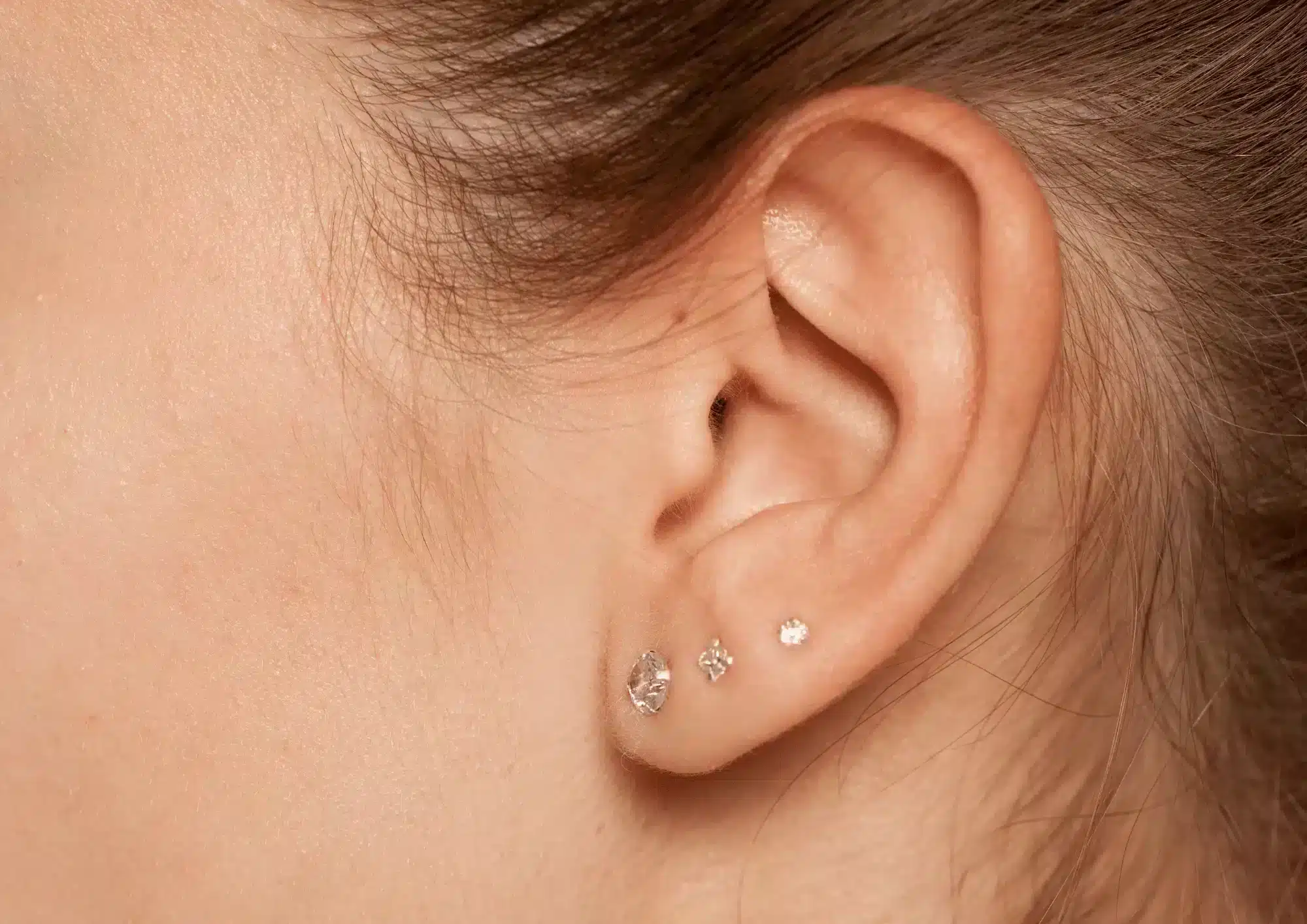
The earlobe is an integral part of the face in an aesthetic sense. For instance, women have often adorned jewelry in this area, with ear piercings being one of the oldest types of body modifications.
Unappealing physical characteristics of the earlobe, such as being too large or having an uneven shape, can become a cause of concern for patients. Additionally, earlobes can be damaged from wearing long, heavy jewelry, which can cause further resulting in undesired aesthetic conditions.
Split earlobe
Over time, patients who wear long or heavy jewelry, especially if suspended from a fine ring, will eventually find that the heavy weight pulling on their piercing stretches, elongates, and sometimes completely pulls through the piercing hole, resulting in a split earlobe. A split earlobe can be incomplete or complete, and different techniques should be used for repair based on the type of damage sustained by the earlobe.
Enlarged ear-piercing hole
The stretching of the ear by wearing special jewelry in the earlobes is a practice that is becoming increasingly popular over the years, particularly among younger people. Stretching of the earlobe is done though a method called gauging, where the earhole piercing is gradually enlarged and stretched by placing different earrings that progressively get larger in size. Using this method, the piercing hole can increase up to 2cm. In order to reduce the hole size, the wearer can gradually decrease the size of the earrings; however, this will not always completely reduce the size of the hole back to its original state.
Anatomy of the ear
In order to treat all of the above concerns, a thorough and detailed understanding of the anatomy is essential. The outer ear consists of the ear canal and the pinna, which is also called the auricle. The pinna is made primarily out of cartilage covered by skin and is visible. The structures that make up the auricle include the antihelix, helix, tragus, lobule (earlobe), and concha. There are two physical types of the lobule: attached, where the earlobe is attached to the side of the cheek, and free, where the lower lobe hangs below the point of attachment to the head.
Blood supply
The outer ear has a rich blood supply since it does not contain cartilage. There are three branches that make up the external carotid artery, the main blood vessel that supplies blood to the area: the occipital artery, anterior auricular branch of superficial temporal artery, and the posterior auricular artery. Each artery has its own corresponding vein that runs together with it. To ensure the flap will still have a supply of blood during the procedure, you should have in-depth knowledge.
External ear sensory innervation
To provide appropriate pain management for the ear, the practitioner should have a deep understanding of nerve anatomy in this area. The ear contains many nerve endings that are a part of four main sensory nerves. These include the:
• Great auricular nerve;
• Auricular branch of vagus nerve;
• Auriculotemporal nerve;
• Lesser occipital nerve.
Treatments
The right technique is instrumental to the success of the treatment. The techniques used to repair a damaged earlobe fall into a few basic categories divided by aesthetic concern, some of which can’t be alleviated by dermal fillers.
Split earlobe repair
To repair a split earlobe, two methods may be employed, with options to reconstruct the piercing hole or not. One can either opt for a straight-line type of repair or a broken line type of repair. These procedures can also be called L-plasty or Z-plasty, respectively, with the letters referring to the shape of the wound after the earlobe is cut. Generally, the broken line of repair is the preferable option because the zig-zag line that is a byproduct of the procedure will not likely produce a notch effect on the free border of the treated earlobe.
If the earlobe split is incomplete, the L-plasty is recommended for its repair, especially against the method of cutting the skin around the edge of the split and stitching, which will actually cause elongation of the earlobe.
Earlobe reduction
Reducing the size of the earlobe can be achieved without notches or scars on the free border by employing the following method:
First, determine the earlobe size your patient wants to achieve, and mark it out with a white marking pen. Draw two lines demarcating the border of excision, first from the tragus to the original marking and the second from the lateral end of the white mark to the tragus. Measure both of these lines and create a third line of the same length as the first two lines, creating a triangle. Excise the earlobe following the lines in place, and then stitch the triangle section in to shorten the second line.
Repairing enlarged piercing holes
For a very large piercing hole, the Y-plasty technique may be employed, whereas the L-plasty is suited for the treatment of large piercing holes, although this will depend on the amount of tissue available.
The procedure approach is outline as below:
• In the case of an L-plasty, draw the lines of excision on the highest point of either side of the cleft. These should be marked before the edges start curving in. This helps to avoid leaving a groove along the repair line.
• Inject an appropriate amount (about 2ml) of local anesthetic into the front and back of the junction of the earlobe and cheek, superficially into the junction, and up to the intertragic notice from the low end of the junction. Suitable anesthetics include xylocaine (2%) with epinephrine or lidocaine (2%).
• Proceed to the apex of the cleft from the free border with Iris scissors or a scalpel. Excise the area according to the markings with extra care taken to ensure that the back and front sides of the earlobe are cut to the same shape. Start at the anterior aspect and insert the resulting flap accurately.
• Use non-absorbable sutures and a vertical mattress technique to stitch the desired shape of the front and back of the earlobe. This technique prevents the scar from rolling inward.
Procedure precautions
The following are some of the precautions you should take when performing an earlobe repair:
• Check the back of the earlobe to determine it is cut to the desired shape. Do this before you start to stich the wound.
• Have the patient sit upright to measure the markings on both ears. This will ensure the shape is similar on both sides. The patient should be made aware that the earlobes may not look identical depending on the condition of either earlobe; however, both sides should still be similar.
• Ensure that all stitches are tightened properly to prevent instance of bleeding.
• To make sure that the stiches do not come loose during the recovery period, advise the patient to refrain from adding stress to the area. To do this they should sleep on their backs (as opposed to their sides) and not play any contact sports or perform any activity that may cause pulling of the ear during the recovery period.
• Your patient should not take any blood-thinning medicines, such as aspirin; vitamins C and E; alcohol, and ibuprofen before the procedure. This is done to minimize the risk of increased bleeding, bruising, and hematoma.
• Triamcinolone (TCN), a glucocorticoid commonly used in these kinds of procedures, is a potential allergen and can cause anaphylactic reactions. Always assess your patient’s risk for hypersensitivity reactions by taking down their medical history, paying particular attention to their history of allergic reactions, especially to steroids.
• Patients prone to keloid or hypertrophic scar formation should be advised that this procedure carries with it the risk of keloid scarring.
• If patients get an infection, they should avoid touching the area and getting the infected area wet for the first 2-3 days after the onset of the infection, if not longer. Patients should also wash their hair prior to their surgery, particularly the morning of, avoid washing their hair for 2-3 days afterwards.
Conclusion
There are many ways to repair a damaged earlobe, including the ones discussed in this article. The techniques outlined are ones that many practitioners find provide optimal results with minimal complication, but its use will depend on the circumstances of each individual case. Treatment must therefore be tailored to the specific individual’s need with relevant factors (e.g. type of repair, anatomy, medical history of the patient) that will impact treatment outcome taken into account.
Aesthetic medicine products are developed and regulated to meet stringent safety and efficacy standards. They are typically administered by trained healthcare professionals such as dermatologists, plastic surgeons, and specialized nurses in clinical settings. These products aim to provide effective solutions for cosmetic enhancement, skin rejuvenation, and overall aesthetic improvement, contributing to both physical appearance and self-confidence.
Key categories of aesthetic medicine products include:
-
Injectables: This category includes products such as dermal fillers, botulinum toxins (e.g., Botox), and collagen stimulators. These injectables are used to smooth wrinkles, add volume, and improve facial contours.
-
Skin Rejuvenation Treatments: Products like chemical peels, microdermabrasion systems, and laser devices are used to improve skin texture, reduce pigmentation irregularities, and enhance overall skin tone.
-
Skincare Products: These include medical-grade cleansers, moisturizers, serums, and topical treatments containing active ingredients like retinoids, antioxidants, and growth factors. They are formulated to address specific skin concerns such as acne, aging, and hyperpigmentation.
-
Hair Restoration Products: Medical treatments and products designed to promote hair growth and treat conditions such as male and female pattern baldness.
-
Body Contouring and Fat Reduction: Devices and products used for non-surgical body sculpting, such as cryolipolysis (cool sculpting) devices and injectable lipolytics.
-
Cosmeceuticals: High-performance skincare products that bridge the gap between cosmetics and pharmaceuticals, often containing potent ingredients with proven clinical benefits.
-
Wound Care and Scar Management: Products like silicone sheets, gels, and advanced wound dressings used to improve healing and reduce the appearance of scars.


